KRF Clinical Practice Guidelines in Keloid Disorder (KRF Guidelines®) Pubic Keloids – Version 1.2019

KRF Clinical Practice Guidelines in Keloid Disorder (KRF Guidelines®) Pubic Keloids – Version 1.2019

Michael H. Tirgan, MD

SUMMARY

Diagnosis

Diagnosis of pubic keloid lesion(s) is based on clinical history as well as the clinical appearance of the skin lesion(s). A biopsy is almost never indicated to establish the diagnosis.

Grouping pubic keloids:

For purposes of this Guideline, pubic keloid lesions are divided into three types. This classification is based on clinical observation and the collective analysis of 65 consecutive patients with pubic keloids. To the author’s knowledge, and after a review of the published literature, there are no published methods to classify public keloids.

  1. Early-Stage lesions presenting as protruding papules, linear, and nodular lesions (< 2 cm – Pubic Stage IA).
  2. Tumoral disease presenting as bulky tumor mass(es).
    • Tumoral – solitary or multiple tumors (2-5 cm – pubic stage IB)
    • Tumoral – semi-massive (5-10 cm – pubic stage IC)
    • Tumoral – massive (>10 cm – pubic stage IIA and higher)
  3. Post C-Section / Supra-Pubic incisional Keloids

Treatment

The choice of treatment for pubic keloids is dependent on the size and the extent of the disease:

  1. Intra-lesional triamcinolone (ILT) shall be the first-line treatment for all early-stage, papular, and linear lesions (see
    KRF Guideline – ILT).
  2. Intra-lesional chemotherapy (ILC) should be considered for all early-stage, papular, and linear lesions that fail to
    respond to ILT (see KRF Guideline – ILC).
  3. Contact-cryotherapy with or without ILT/ILC is the preferred and the primary method of destruction of all nodular
    and tumoral keloid lesions (see KRF Guideline – Cryotherapy).

Rationale for the use of cryotherapy:
a. Cryotherapy is an effective method of treatment for protruding and bulky keloids.
b. As opposed to surgery, cryotherapy does not cause the worsening of keloids.
c. As opposed to surgery, radiation therapy is unnecessary after cryotherapy [1-4].

Treatments to avoid:

Surgery shall not be used in the treatment of early stage pubic keloids. The disease process in most patients is multifocal and progressive. Furthermore, surgical intervention is a known cause for the worsening of keloids [1, 2], which is also well documented in several cases presented in this Guideline.

Radiation therapy shall not be used in the treatment of pubic keloids. This intervention may induce neoplastic transformation of irradiated tissues, in particular in proximity to the pelvis [3,4].

Lasers shall not be used in the treatment of pubic keloids. This intervention may result in the worsening of keloids [5]. Overview:

This KRF Guideline was developed with the aim to provide:

  1. A general discussion of pubic keloids.
  2. The natural history of pubic keloids.
  3. The classification system for pubic keloids.
  4. Recommendations for treatment and follow up.

INTRODUCTION

Keloid involvement of the pubic skin is fairly uncommon and occurs in approximately 6% of patients with keloid disorder. In a recent analysis of data from 1,088 consecutive patients seen by the author in his keloid specialty practice, there were 65 (6.0%) patients with keloid involvement of the pubic skin. This pattern of presentation of keloid disorder is highly gender and race specific. Among this cohort of patients, 56 (86%) were female and 9 (14%) male; 54 (82%) were African-American, 5 (8%) were Asian, 3 (5%) were Caucasian, and 3 (5%) were Hispanic.

Clinical presentation

The demographics and patterns of presentation of the diseaseamong the cohort of 65 consecutive patients with pubic keloids is summarized in Table 1. Clinical presentation of pubic keloids is distinctly race specific, with the tumoral and more advanced forms of the disease almost exclusively seen among African-Americans. Fiftyfour (82%) of patients presented with tumoral form of the disease with most (25 patients, 39%) presetting with massive tumoral pubic keloids, measuring 10 centimeters or larger in total diameter.

Table 1. Demographics of patients and morphology of pubic keloids.

Demographics of patients morphology

The primary triggering factors that prompt formation of early-stage pubic keloids have never been properly studied. Injury during pubic hair removal process, or ingrown hair may be the triggering factors for development of early stage disease. Surgery to remove a small pubic keloid and a C-Section procedure are common triggering factors for the formation of much larger keloids.

Involvement of the pubic skin as the only site of keloid disorder was seen in about one third of the patients (20 cases, 31%). Most patients (45 cases, 69%) in addition to their pubic keloids, however, have keloid lesions elsewhere on their skin.

What stands out in the clinical picture and patterns of presentation of pubic keloids among these 65 individuals is that among Asians, Hispanics, and Caucasians, pubic keloids present in a much more limited fashion. However, despite the small sample size, among 11 patients who were not African-American, only one Asian female patient had a tumoral keloid (measuring approximately 2 cm) in the pubic area (shown in Figure 8) and one other Asian female patient had a large patch of flat keloids in her pubic area (shown in Figure 20). The other nine patients in this cohort of 11 had very limited involvement of their pubic area, with the disease appearing as small papules or nodules.

The most important goals of treatment are:

a. To intervene very early and to bring the earlystage lesions under control with a combination of intra-lesional triamcinolone (ILT) / intra-lesional chemotherapy (ILC) and cryotherapy.

b. To avoid surgery in all patients. Surgical removal of pubic keloids – a dynamic and multifocal disease – is an inherently flawed approach that exposes patients to the unnecessary risk of developing massive tumoral keloids.

c. To bring the triggering factor(s) under control. Patients should be educated about potential risk of injury to their pubic skin from mechanical methods of pubic hair removal such as waxing or shaving using blades. These methods can not only directly damage the skin, but also can lead to the formation of ingrown hair, which in turn can result in folliculitis that can trigger keloid formation. These two traumatic methods of pubic hair removal should be avoided in all patients as they can increase the chance of injury to the pubic skin. All patients with pubic keloids should consider using electric trimmers to trim their pubic hair as this method reduces the chances of injuring the pubic skin. Laser hair removal may also induce injury to the pubic skin, thereby triggering new keloid formation.

Early-stage Pubic Keloids

Pubic keloids at their earliest stages appear in three distinct manners:

  1. Protruding papule(s) that often form in the pubic area (Figure 1). If left untreated, these lesions can grow to become nodular keloids.
  2. Linear lesions. If left untreated, these lesions can grow to form thicker linear lesions (Figures 2, 3).
  3. Nodular lesions. If left untreated, these lesions can grow to form tumoral lesions (Figures 2, 3).

pubic keloid

Figure 1. Early-stage protruding pubic keloid papule in a 26-yearold African-American female who also had a large tumoral anterior chest keloid present for close to 10 years.

pubic keloid

Figure 2. Early-stage linear pubic keloid in a 34-year-old Asian female with a 10-year history of keloid disorder involving the anterior chest and pubic areas.

pubic keloid

Figure 3. Early-stage nodular pubic keloid in a 34-year-old African-American female with a six-year history of solitary pubic keloid as the only site of the disease involvement.

Analysis of the clinical data of the 65 patients with pubic keloids suggests that the disease entity at its early stages may take on a variable course of progression. Careful observation and analysis of the clinical data identifies two biologically distinct patterns of progression of the disease in patients with early stage pubic keloids.

  1. Group A. Patients who only develop one or a few small keloid lesions over an extended period of time (five to 10 years) and their disease follows a very slow pace. This is perhaps the case for some Asian as well as Caucasian women with keloid disorder. Figure 2 depicts a young Asian female who presented to the author in February 2013. Over the span of 10 years, she had developed only two small lesions, one in pubic area (Figure 2) and one on her anterior chest skin. Figure 4 depicts the same lesion, but in July 2016, almost three and a half years later, after several attempts at treatment. While undergoing treatment for over three years, this patient did not develop any new keloid lesions.
  2. Group B: Patients for whom the disease progresses at a continuous pace and over time, new lesions form and existing lesions grow in size. This is perhaps the case for most African-American patients with keloid disorder, especially those undergoing keloid removal surgery.

pubic keloid

Figure 4. The same patient as in Figure 1 in July 2016. Note the persistence and discoloration of the original lesions yet the absence of new keloid formation in a span of 3.5 years.

Treatment

There are two distinct goals for treatment of patients with early-stage pubic keloids:

  1. To induce remission.
  2. To prevent progression/worsening.

It is of utmost importance to manage early-stage keloids in a manner that prevents further growth and the formation of large and tumoral keloids. Both the treating physician and the patient must be cognizant that the disorder is multifocal with a dynamic pathophysiology, and while the triggering factors are still active, with the passage of time the existing lesions will continue to grow in size. Most patients, in particular African-Americans, are destined to develop new lesions either in the pubic area or other (distant) parts of their skin. It is naïve to think that the keloid process is static or is limited to the pubic skin such that it can be surgically treated. All keloid patients must be treated in accordance with a longterm treatment and follow-up plan. Patients also need to be adequately educated about their illness and its biology.

ILT injection shall be the first-line treatment for all earlystage papular pubic keloid lesions. The lesions must be followed carefully and ILT shall be continued on a regular basis to achieve maximum response. It is of equal importance to identify the triggering factors and avoid using blades or waxing for hair removal.

ILC should be considered early on for all lesions that fail to respond to ILT.

Contact Cryotherapy shall be the first line of treatment for all nodular pubic keloids. Any residual keloid tissue that may remain after cryotherapy shall be treated with ILT and/or ILC.

pubic keloid

Figure 5. Solitary pubic keloid tumor in a 34-year-old African-American female with a 13- year history of pubic keloids. This recurrent keloid grew at the site of a prior pubic keloid that was removed surgically.

pubic keloid

Figure 6. A 27-year-old Hispanic female presented in September 2012 with multiple papular/linear keloids and a tumoral pubic keloid. The tumoral lesion was treated with cryotherapy during the same visit.

pubic keloid

Figure 7. The same patient in March 2013, following recovery from cryotherapy. Note the near total debulking of the tumoral lesion.

An untreated or poorly treated early stage pubic keloid can eventually grow in size and expand three-dimensionally to form a tumor. For purposes of this Guideline, tumoral pubic keloid lesions are divided into three types:

  • Tumoral – solitary or multiple tumors (2-5 cm – pubic stage IB)
  • Tumoral – semi-massive (5-10 cm – pubic stage IC)
  • Tumoral – massive (>10 cm – pubic stage IIA and higher)

Tumoral pubic keloids are almost exclusively seen among African Americans. Among 53 patients with tumoral pubic keloids, 49 (95%) were African-American.

Table 2. Subtypes of tumoral pubic keloids

pubickeloid

Most patients with tumoral keloids have a history of prior keloid removal surgery. The injury from surgery to remove a small pubic keloid triggers the formation of much larger tumoral keloids. Therapeutic interventions shall aim at preventing tumoral keloids by avoiding surgery in the management of early-stage pubic keloids.

Treatment

Cryotherapy is the treatment of choice for most tumoral pubic keloids as these tumors respond very well to cryotherapy and can be significantly debulked with one or two treatments. Cryotherapy often results in a durable debulking as shown in several cases in this Guideline.

Case Study 1

A 34-year-old Asian female presented in June 2014, with two tumoral keloids, one on her pubic area (Figure 8) and one on her anterior chest area. She reported that the chest keloid had been present for about 15 years yet the pubic keloid was a more recent development, present for only three years. She recalled that both keloids had formed following an inflammatory skin lesion, what she described as a “pimple,” and had continued to grow at a constant rate. She had never received any treatment for her pubic keloid. The pubic keloid was treated with contact cryotherapy (Figure 9).

pubic keloid

Figure 8. A 34-year-old Asian female with a previously untreated tumoral pubic keloid (June 2015).

pubic keloid

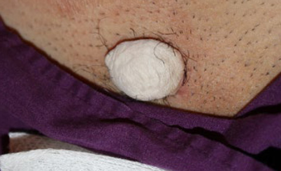
Figure 9. The same patient immediately after application of cryotherapy (June 2015).

pubic keloid

Figure 10. The same patient after recovery from cryotherapy (January 2016). Significant debulking of the keloid was achieved with only one cycle of treatment. The base of this keloid needs continued treatment as there is a visible residual disease.

Multi-Focal Tumoral Disease

Similar to the keloid process in other regions of skin, the pubic keloid process, especially among African-American females, is often multi-focal and tumoral. Figure 11 depicts multiple tumoral keloids in a 45-year-old African-American female, who at the same time had several large chest, shoulder, and upper arm keloids. Multi-focal tumoral pubic keloids are the most frequently seen type of keloids among African American females.

pubic keloid

Figure 11. A 45-year-old African-American female with multiple tumoral pubic keloids.

pubic keloid

Figure 12. A 40-year-old African-American female with multiple tumoral pubic keloids. This patient also had multiple large keloids on her chest and shoulders.

pubic keloid

Figure 13. A 37-year-old African-American male with multiple tumoral pubic keloids. This patient presented with several other large keloids on his neck, face, and chest.

Negative Impact of Keloid Removal Surgery

With the passage of time, most tumoral pubic keloids continue to grow and merge to form large tumoral patches. Treating pubic keloids can be quite challenging, no matter how small the lesions may be. Although the option of surgery followed by radiation therapy might result in reasonable outcomes for some patients [6], the risk of recurrence and exposure of the pelvic area to radiation in a young person cannot be ignored. As exemplified in case study 1 presented earlier in this Guidance and case study 2 described below, reasonable outcomes can be achieved with cryotherapy. It is also reasonable to conclude that in contrast to surgery, early implementation of non-surgical treatments on early-stage and small pubic keloids has the potential to result in better long-term outcomes.

Keloid removal surgery is a significant risk factor for the development of large tumoral or superficially spreading keloids (Figure 19). As seen in this Guideline, the injury from surgery to remove a small pubic keloid can trigger formation of much larger tumoral keloids.

Case Study 2

A 22-year-old African American female presented in October 2014, with multiple tumoral keloids on her ears, chest, shoulders, abdomen, and pubic area. Many of her keloids had been previously excised surgically yet each one had regrown to become a much larger keloid. Figure 15 depicts the status of this patient’s pubic keloid at presentation.

I am doing fine. I have been waiting for your directives about the other books.

Figure 16 shows the results there were achieved after recovery from one cycle of cryotherapy in February 2015.

pubic keloid

Figure 14. A 45-year-old African American male with multifocal pubic keloids. This patient has been struggling with keloid disorder involving several areas of his skin since the age of 19.

pubic keloid

Figure 15. A 22-year-old African-American female with multiple tumoral pubic keloids (October 2014). All keloids shown were treated with cryotherapy in one session.

pubic keloid

Figure 16. The same patient in February 2015, after recovery from one cycle of cryotherapy. Note the significant reduction in the mass of all treated keloids. The base of each treated keloid must be treated with ILC and ILT to reduce the chances of recurrence.

Case Study 3

A 33-year-old African-American female presented to the author in June 2013, with multiple tumoral pubic keloids (Figure 17). Her struggles with keloid disorder started when she was 13 years of age. Over the next two decades, similar to the previous patient, she underwent multiple surgeries to remove keloids from her pubic area as well as from her ears. Unfortunately, these interventions caused worsening of her keloids, both on her ears as well as her pubic area. Most of the bulky pubic keloids were treated with cryotherapy. Unwilling to undergo anymore surgeries, she sought treatment with cryotherapy. Figure 18 depicts the results achieved with two cycles of cryotherapy.

Over time, and importantly triggered with repeated surgeries, pubic keloids tend to regrow and involve larger areas of the skin. Figures 19-22 depict other cases treated with repeated and indiscriminate surgeries, which led to very unfortunate outcomes. Treatment of such advanced and recurrent pubic keloids is quite challenging. The key message here is that pubic keloid removal surgery is the most important risk factor for the formation of large, semi-massive and massive pubic keloids. The only way that we can avoid formation of these keloids is to refrain from performing surgery to remove early-stage pubic keloids.

pubic keloid

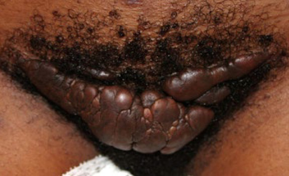
Figure 17. A 33-year-old African-American female with multiple tumoral pubic keloids after undergoing several attempts at surgical removal of her pubic keloids (June 2013).

pubic keloid

Figure 18. The same patient in January 2014, after recovery from two cycles of cryotherapy. Note the reduction in the mass of all treated keloids. The remaining keloids still must be treated with more cryotherapy and then with ILC and ILT.

pubic keloid

Figure 19. A 23-year-old African-American female with multiple tumoral pubic keloids after undergoing several attempts at surgical removal of her pubic keloids.

pubic keloid

Figure 20. A 24-year-old Asian female with extensive keloids of the abdominal wall and pubic areas. This patient had undergone a renal transplant, which required several abdominal and pubic surgeries. Most of her keloids were formed at the sites of the skin injury from surgical procedures.

Massive Pubic Keloids

Massive pubic keloids ( >10 centimeters in diameter, Pubic Stage IIA and higher) are almost exclusively seen in African Americans (22/23) and mostly those who have undergone surgery to remove a previous, smaller keloid (21/23). These patients all have other keloid lesions elsewhere on their skin (23/23).

Treating these patients is quite challenging as most have already been treated with all available therapeutic interventions. Ideally, these patients need to be treated with a systemic treatment, i.e. a drug that can be safely administered orally or intravenously. Unfortunately, there are no systemic treatments available at the current time and none even on the horizon.

pubic keloid

Figure 21. A 28-year-old female with extensive tumoral pubic keloids. Her struggle with pubic keloids started at age of 17. Over the years, she underwent three separate surgeries with each making her keloids worse.

pubic keloid

Figure 22. A 28-year-old African American female with massive tumoral involvement of the pubic area (May 2014).

pubic keloid

Figure 23. The same patient after recovery from cryotherapy for two portions of her keloid. Note that near total removal of keloid masses is achievable with cryotherapy (September 2015).

pubic keloid

Figure 24. The same patient after recovery from cryotherapy for other portions of this keloid. Although not ideal, significant debulking was achieved with the reduction in the overall mass of this keloid (June 2017). This patient still needs several more rounds of treatment to remove the rest of the bulky keloids from her pubic skin. Note the durable results of the treatment in areas treated in 2013.

Limited Role of surgery:

Surgery, using a scalpel or a laser device, should never be used to remove early-stage, nodular, multi-nodular, or even semi-massive tumoral pubic keloids. Surgery is the triggering factor for the formation and development of much larger keloids.

Surgery may only be considered in cases of massive pubic keloids and only performed in coordination with a specialist physician familiar with keloid disorder, who can administer proper adjuvant medical treatments, including ILC, in an attempt to prevent a postoperative recurrence.

When contemplating surgery for patients with massive keloids, one must be reminded that the lesion that is to be removed was triggered by previous surgical interventions to remove much smaller lesions.

Post C-Section / Suprapubic Incision Line Keloids

The occurrence of post-operative keloids is a challenging complication of suprapubic incisions in young African American females. The injury from surgery triggers this keloidal wound healing reaction in genetically prone patients. Over time, the surgical wound healing is complicated with a keloid reaction, leading to the formation of keloidal tissue along the lines of the C-Section and other sites injured during the surgery.

Although seen and reported among the Asian population [6], in the United States these keloids are almost exclusively seen in African American female patients. Among the cohort of 65 patients seen by the author, there were 20 women with C-Section keloids, all of whom were African American. To the author’s knowledge, there are no reports of the true incidence of this complication after supra-pubic incisions.

Treatment of these keloids, especially when they present at advanced stages, is rather difficult. The author recommends early and aggressive intervention: when possible with ILT and ILC at the earliest signs of keloidal transformation of the C-Section wounds in women who are not breastfeeding. ILT and ILC shall not be used when women are nursing. In addition, many young women who have just given birth are too busy attending to their newborn and taking care of their families, as a result, they see no choice but to forgo treatment of their incisional keloid.

Often times, these keloids are excised during a subsequent C-Section such that yet again the cycle repeats often leading to the formation of a new keloid.

Cryotherapy is the treatment of choice for tumoral keloid formation on the C-Section incision line, as it is the safest treatment option.

Case Study 5

A 28-year-old African-American female presented in June 2013, with multiple keloid lesions involving her neck, scalp, chest, and shoulders. She had recently undergone a C-Section and her incision was healing well with minimal signs of linear hypertrophic skin reaction (Figure 25).

This patient was seen several times for treatment of her neck keloids. In March 2015, the patient reported that her suprapubic incision had turned into a keloid (Figure 26). To the extent known to the author, this patient had not received any past treatment for this lesion.

This case illustrates several facts about keloid disorder. Most importantly, the case shows that the disease process is multifocal and is triggered by an injury to the skin. Furthermore, the rate of growth and development of this pubic keloid is established to be less than two years in a patient with fairly advanced keloid disorder, with numerous lesions elsewhere on her skin. Figures 27 and 28 depict two more cases of C-Section keloid disorder in African-American females.

pubic keloid

Figure 26. A 28-year-old African-American female with a recent history of a suprapubic incision (June 2013).

pubic keloid

Figure 27. Same patient, March 2015. Note the formation of a thick linear keloid along the previous supra-pubic incision line.

pubic keloid

Figure 28. A 35-year-old female with a tumoral post C-Section pubic keloid. This patient had three prior C-Sections, all of which resulted in the formation of pubic keloids removed during the next C-Section procedure. The last C-Section was performed approximately 2.5 years prior to this presentation. Other than this keloid, this patient had only small keloid lesions on her shoulder.

pubic keloid

Figure 29. A 25-year-old African-American female with post-C-Section keloid and a massive tumoral pubic keloid that had developed after the surgical removal of a smaller tumoral pubic keloid at the same site. Presence of pre-existing pubic keloids is an indicator for the development of C-Section keloids.